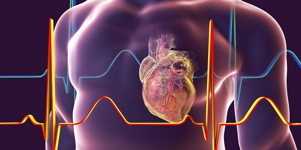
Different types of electrodes are used depending on the specific test or procedure being performed. In general, electrodes stick temporarily to the skin's surface and are connected by wires to an electrocardiography (ECG) machine. The ECG machine then analyzes and records the heart's rhythms and electrical signals.
Types of Electrodes
There are several main types of Cardiology Electrodes used in cardiology. Surface electrodes are the most common and are used to perform basic ECG tests. Around 10 small, sticky pads known as surface electrodes are placed on specific points on the chest, arms, and legs. These detect the heart's electrical signals and transmit them to the ECG machine for analysis. Holter monitor electrodes involve wearing a portable ECG monitor for 24-48 hours using electrodes attached with adhesive tape or elastic belts. This allows doctors to monitor heart rhythm over a longer period to detect any irregularities. Pacemaker electrodes are thin insulated wires with a small electrode at the tip that are implanted through a vein into the heart muscle or chambers. They deliver electrical pulses from an implantable cardioverter defibrillator (ICD) or pacemaker to regulate abnormal heartbeats. Catheter electrodes are located at the tip of specialized catheters inserted into the heart through blood vessels during cardiac catheterization, angiograms, or ablations. They map the heart's electrical activity in more detail.
Surface Electrocardiography
Surface ECG remains the most common initial test to evaluate the heart's electrical activity and rhythm. Small sticker electrodes are placed on both arms and legs, as well as precise intercostal spaces on the chest. This setup is known as a 12-lead ECG as it involves 10 electrodes that produce 12 different views of the heart's activity. The heart's rhythms and electrical conduction pathways between the upper (atria) and lower (ventricles) chambers can be analyzed from these multiple angles. Abnormalities may indicate conditions like arrhythmias, heart attacks, enlarged heart chambers, or other issues. A basic ECG is noninvasive, low-cost, and provides doctors valuable diagnostic information quickly at the point of care.
Holter Monitoring Electrodes
While a single ECG provides a snapshot in time, Holter monitoring uses electrodes attached continuously for 24-48 hours to track any irregular heartbeats or changes in rhythm over an extended period. Small sticky electrodes are connected by wires to a portable ECG recorder worn on a belt or harness. As the patient goes about their daily activities, the system records the heart's electrical signals. Afterward, the recorded data is analyzed by specialized software looking for any abnormalities. Conditions like arrhythmias may only occur intermittently and could potentially be missed with a standard ECG. Continuous monitoring via Holter helps improve detection and diagnosis for various arrhythmia types including atrial fibrillation.
Cardiac Catheterization Electrodes
When more detailed anatomical and electrical mapping of the heart is needed, cardiac catheterization procedures use specialized catheter electrodes. A thin, flexible tube called a catheter is inserted through a blood vessel in the leg, guided to the heart chambers under imaging guidance. Miniature electrode tips at the catheter's end allow detailed localization and recording of electrical signals from inside the heart musculature. This can identify areas of abnormal conduction causing arrhythmias. Catheter ablation then uses radiofrequency energy delivered through the catheter electrodes to scar and isolate problematic heart tissue, curing some arrhythmia types permanently. Mapping data also helps guide device implantation for pacemakers and ICDs. Catheterization provides an important next step after noninvasive tests when further cardiac examination and treatment are indicated.
Implantable Cardiac Device Electrodes
For individuals with irregular heartbeats not controlled by medications, implantable cardiac devices deliver therapy via thin electrode wires placed within the heart. During a procedure, electrode catheters are inserted through veins in the groin and threaded into the heart chambers under fluoroscopy. The electrode tip adheres gently to the endocardial surface via a helical coil. Other electrodes may be placed outside the heart in major veins. Once optimally positioned, these pacing leads deliver regulated electrical pulses from devices like pacemakers and ICDs. Implantable electrodes have contributed greatly to managing abnormal rhythms long-term through calibrated impulse delivery from internally implanted generators. Continuous monitoring also allows prompt response if dangerous fast rhythms arise with defibrillation shock therapy.
In summary, different types of electrodes play critical diagnostic and therapeutic roles through cardiology. From noninvasive surface ECGs to intricate mapping within heart chambers, electrodes allow clinicians to both detect and treat abnormalities of cardiac electrical conduction. Continuous technological advances will surely lead to improved capabilities, yet basic surface ECGs still remain the first-line test when evaluating patients' heart function. Proper knowledge and use of various electrode modalities helps cardiologists optimize cardiac care through multi-faceted assessment, diagnosis and management of heart conditions.
Get more insights on This Topic- Cardiology Electrodes


